Most people understand that stress causes hair loss. What they do not understand — and what the clinical literature has been articulating with increasing precision since the early 2000s — is that chronic stress does not merely cause temporary shedding. Sustained psychological stress lasting two or more years can permanently accelerate androgenetic alopecia in genetically predisposed individuals, effectively compressing a decade of follicle miniaturisation into a two-to-three year window. For KL professionals navigating sustained workplace pressure, this distinction between temporary and permanent is not academic. It determines whether intervention now preserves options that will no longer exist in three years.
The mechanism is not a single pathway. It is the convergence of three independent biological processes that, under chronic stress, compound each other.
Acute vs Chronic: Two Fundamentally Different Conditions
Acute telogen effluvium — the kind triggered by a severe illness, surgery, childbirth, or an acute high-stress period — is a reversible event. Under acute physiological stress, the HPA axis elevates cortisol sharply, CRH is expressed in the scalp, and a significant proportion of anagen follicles are abruptly shifted into telogen. Shedding follows 6–12 weeks later. Once the trigger resolves and cortisol normalises, follicles reenter anagen and density is substantially restored within 6–12 months.
Chronic stress operates on an entirely different timescale and produces structurally different outcomes. With sustained HPA activation lasting months to years, cortisol elevation is not acute and then resolved — it is persistent and baseline-shifted. The downstream effects accumulate without the recovery phase that makes acute telogen effluvium reversible.
| Stress Type | Duration | Cortisol Profile | Hair Loss Pattern | Reversibility | |---|---|---|---|---| | Acute (illness, surgery) | Weeks | Sharp spike, then normalises | Telogen effluvium, diffuse shed | High — most density recovers | | Subacute (project crunch, exam) | 1–3 months | Elevated, then gradually normalises | Moderate shedding, delayed | Moderate — partial recovery | | Chronic (sustained workplace) | 2+ years | Persistently elevated baseline | Diffuse thinning + accelerated AGA | Low if AGA progression has locked in | | Chronic + Genetic AGA | 2+ years | Persistently elevated + DHT sensitivity | Rapid pattern thinning | Minimal without early intervention |
The Cortisol–DHT Sensitisation Pathway
Chronic cortisol elevation does not directly cause androgenetic alopecia. Its role is more insidious: it sensitises follicles to dihydrotestosterone (DHT) by upregulating androgen receptor expression in dermal papilla cells. A follicle that under normal conditions would be minimally responsive to circulating DHT becomes progressively more reactive under sustained cortisol load.
In parallel, chronic cortisol suppresses the Wnt/β-catenin signalling pathway — the primary molecular switch for anagen phase initiation. The result is a follicle that is both more sensitive to the miniaturising signal of DHT and less capable of mounting the regenerative response of anagen entry. These two effects are synergistic. In a person with genetic predisposition to AGA — meaning they carry androgen-sensitive follicles in the crown and frontal hairline — two years of sustained cortisol elevation can produce follicle miniaturisation that would otherwise take a decade under normal DHT exposure.
This explains a clinical pattern that is increasingly common in KL: professionals in their late 20s and early 30s presenting with hairline recession and crown thinning that appears "too early." The genetics were always there. The stress timeline compressed the schedule.
Substance P and the Neurogenic Inflammation Loop
The Arck et al. 2006 neuroimmunology framework introduced a second pathway that operates independently of cortisol: neurogenic inflammation via Substance P. Under psychological stress, cutaneous sensory nerve fibres release Substance P directly into the perifollicular dermis. Substance P activates mast cells — which then degranulate, releasing histamine, proteases, and pro-inflammatory cytokines including TNF-α and IL-1β — within millimetres of the follicle bulge region.
This perifollicular inflammatory environment damages the follicular stem cell niche housed in the bulge. Unlike the dermal papilla, which can recover from temporary stress, the bulge stem cell population has limited regenerative reserve. Repeated Substance P-driven mast cell activation at the bulge — the kind that accumulates over years of chronic stress — can produce irreversible fibrosis in the perifollicular connective tissue sheath. Once this fibrosis is established, the follicle cannot re-enter productive anagen regardless of hormonal environment. This is the permanent endpoint.
Why Topical Treatments Fail in Chronic Stress Conditions
The clinical frustration reported by many KL patients is this: they are using evidence-based topical treatments — minoxidil, peptide serums, caffeine formulations — without meaningful response. The mechanism is now understood. Minoxidil's primary action is vasodilation of the dermal papilla microvasculature, improving oxygen and nutrient delivery to stimulate anagen. It is effective when the follicle retains the biological capacity to respond to that stimulus.
Under chronic cortisol elevation, the Wnt/β-catenin suppression means the follicle's anagen initiation machinery is pharmacologically inhibited at a point upstream of where minoxidil acts. The vascular stimulus arrives; the molecular response is blunted. Similarly, sustained perifollicular inflammation creates a local environment where growth factor signalling is competitively suppressed by pro-inflammatory cytokines. The treatment is reaching a follicle in a state that prevents it from responding.
Treating the HPA axis is therefore not complementary to follicle treatment — it is prerequisite.
The TTE Approach: Regulating the Upstream Driver
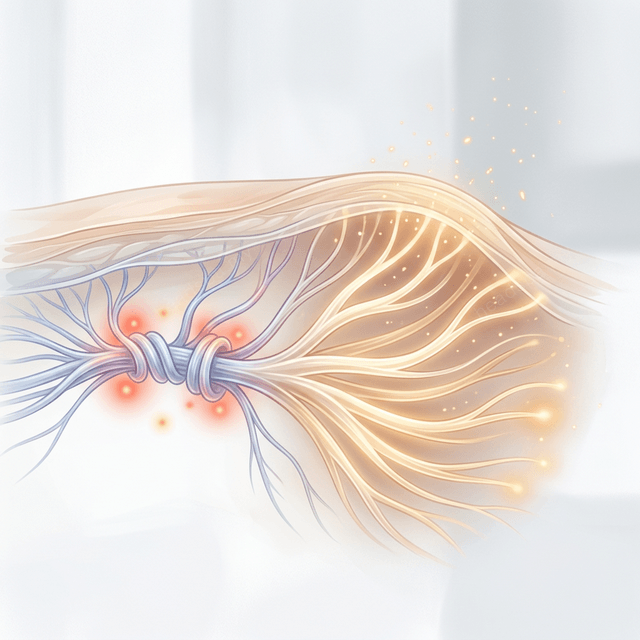
The [TTE Sleep Healing Headspa](/sleep-healing) protocol is structured around a documented clinical observation: vagus nerve stimulation produces measurable HPA axis downregulation, promoting relaxation and supporting cortisol regulation following consistent parasympathetic activation protocols. The treatment's craniosacral-influenced pressure work, thermal gradient sequencing, and acoustic environment are not separate modalities — they are coordinated inputs to the autonomic nervous system.
Understanding the [vagus nerve's role in scalp health](/blog/vagus-nerve-scalp-connection) clarifies why this is the logical upstream intervention. The [cortisol–follicle pathway](/blog/cortisol-follicle-pathway) details the downstream consequences when that intervention does not occur. For KL professionals who have been experiencing sustained workplace stress for more than 12 months, the question is not whether their scalp is being affected — the biology is unambiguous on this point. The question is whether intervention now occurs before the irreversibility threshold is crossed.
The intervention window matters. [Book a clinical assessment at TTE KL](/headspa-kl) or [TTE JB](/headspa-jb) to evaluate current follicle status and stress-load history.
---
FAQ
Q: How do I know if my hair loss is stress-related or genetic? A: The distinction requires clinical assessment. Indicators of stress-driven acceleration include: rapid onset over 12–24 months, history of a significant stress period preceding the loss, diffuse thinning across the crown rather than purely at the temples, and elevated perceived stress score. A trichoscopy assessment can evaluate the proportion of miniaturised versus active follicles and indicate whether the loss pattern is consistent with accelerated AGA or primary telogen effluvium.
Q: I've had high stress for 3 years. Is my hair loss already permanent? A: Not necessarily — and this is a critical distinction. Follicle miniaturisation is a spectrum. Early miniaturisation (reduced shaft diameter, shortened anagen) is often partially reversible with combined cortisol reduction and appropriate follicle-stimulating protocols. Established fibrosis of the perifollicular sheath represents the irreversibility threshold. Clinical assessment of your specific follicle status determines what recovery is still possible. Earlier intervention preserves more options.
Q: Can stress management alone stop my hair loss? A: Stress reduction is the most important lever for stopping the progression of stress-accelerated AGA — but it must be combined with appropriate topical or systemic follicle support. Reducing cortisol restores Wnt/β-catenin signalling capacity and reduces DHT sensitisation; it does not independently reverse existing miniaturisation. The combined protocol — nervous system regulation plus follicle-stimulating treatment — produces outcomes neither achieves alone.
Q: Is there a specific cortisol level that indicates danger for follicle health? A: Clinical research identifies evening cortisol elevation as the most follicle-relevant marker — specifically, failure of the normal cortisol nadir between 10pm and 2am. Morning cortisol is less predictive of follicle damage than the loss of evening suppression. Salivary cortisol testing at 11pm provides a practical assessment of HPA axis dysregulation in a clinical context.
Q: SOCSO data mentions burnout — does burnout specifically accelerate hair loss faster than regular stress? A: Burnout is characterised by HPA axis exhaustion, which paradoxically can present with flattened cortisol (low morning cortisol, low evening cortisol) rather than the elevated profile of acute stress. This late-stage pattern may be less immediately damaging to follicles than the sustained elevation phase that precedes it — but the accumulated perifollicular inflammation and prior Wnt suppression from years of cortisol overload create lasting structural effects. The damage in burnout cases is often the accumulated legacy of the sustained-stress phase.